Temporal Bone Surgery Drill Simulator
Otolaryngology / Temporal Bone Surgery / Skills Training
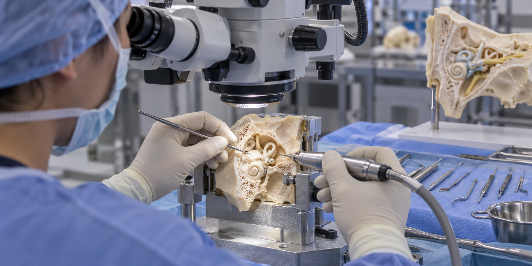
The Challenge
Temporal-bone drilling is one of the most challenging parts of ENT training. Junior residents must work under a microscope in a very small, complex three-dimensional space while controlling a surgical drill and suction.
Critical structures including the facial nerve, inner ear, carotid artery, dura, and major venous sinuses lie close to the field. Residents must recognize anatomical landmarks, maintain orientation, choose a drilling strategy, coordinate their hands, control depth and angle, and know when they are approaching structures that must not be injured.
Why Current Tools Fall Short
Training combines supervised operating-room experience, temporal-bone dissection courses, and laboratory practice. Operating-room time is limited, and teaching during a real procedure is difficult.
Cadaveric temporal bones are realistic but limited by cost, availability, ethical and cultural considerations, regulation, specialized facilities, single-use specimens, and variable anatomy that makes a standardized progression difficult. 3D-printed models provide more accessible and standardized anatomy, but are usually single-use for drilling and do not fully reproduce the tactile properties of bone.
Virtual simulators allow repeated cases and different anatomical configurations, and some add haptic resistance. The remaining opportunity is to combine this repeatability with progression, challenges, immediate feedback, scoring, mastery, and meaningful consequences.
Possible Directions
One possible direction is a virtual drilling environment in which a resident begins from recognizable landmarks, chooses a safe strategy, maintains orientation, and exposes a target structure while preserving protected anatomy.
The experience could represent the drill in one hand and suction in the other; vary bone density, anatomy, and case difficulty; warn when the drill approaches the facial nerve, inner ear, carotid artery, dura, or venous sinuses; and allow immediate reset after a mistake. VR could support the three-dimensional view, while optional haptics could reproduce different tissue resistance.
Progressive cases could move from landmark identification and a simple exposure to unfamiliar anatomy and more complex drilling decisions. The same framework could later support other technically demanding surgical procedures.
What Success Could Look Like
- Better recognition of landmarks and a clearer three-dimensional mental model.
- Appropriate drilling strategy and maintained orientation.
- Accurate target exposure with fewer contacts with protected structures.
- More efficient drilling, less unnecessary bone removal, and successful completion of increasingly complex cases.
- Improvement across repeated attempts and better preparation for supervised surgery.